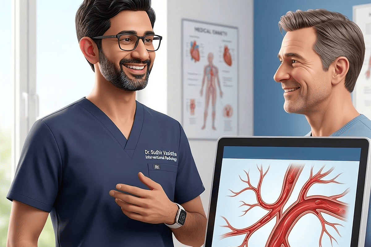
Lungs & Chest (Thoracic) Interventional Procedures | Interventional Lymphatic Procedures
Embolization | Age: Adults | Geriatrics
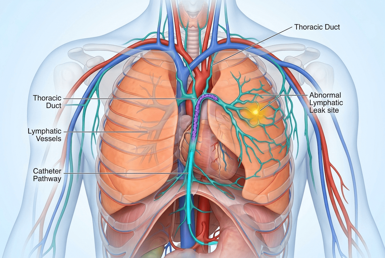
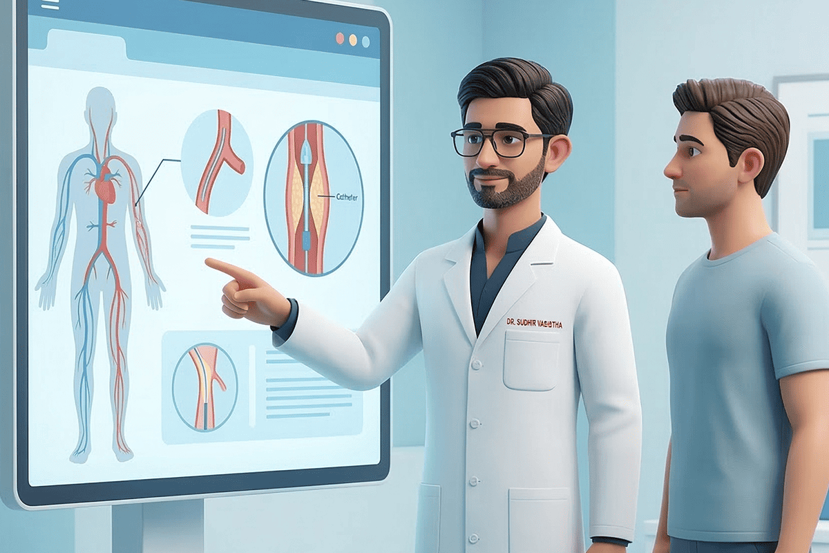
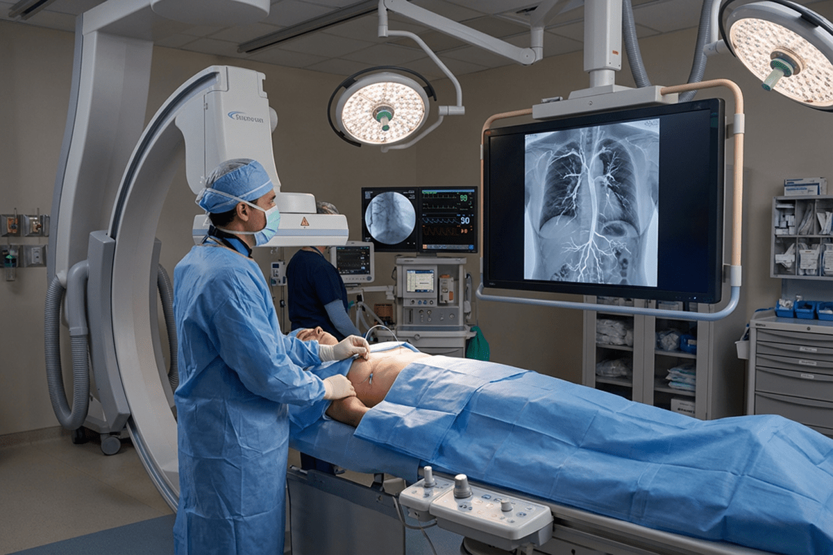
Lymphangiography is a specialized, minimally invasive imaging procedure used to visualize the lymphatic system and identify abnormal lymphatic leaks, blockages, or malformations. Using advanced image guidance, contrast material is introduced into lymphatic vessels to map lymph flow in detail. When a leak or abnormal pathway is identified, thoracic duct embolization can be performed to seal the affected lymphatic vessel and stop ongoing fluid leakage.
Thoracic duct embolization is commonly used to treat complex lymphatic disorders such as chylothorax and postoperative lymphatic leaks. These interventional lymphatic procedures provide both diagnostic clarity and effective treatment, often eliminating the need for open surgery and significantly improving patient outcomes.
Internal links: Lungs & Chest (Thoracic) Interventional Procedures, Interventional Lymphatic Procedures, Embolization
Lymphangiography and thoracic duct embolization are advanced but well-established procedures when performed by experienced interventional radiologists. Potential risks include infection, bleeding, contrast reaction, or incomplete leak closure. These risks are uncommon and carefully managed through detailed imaging, expert technique, and close follow-up care.
Lymphangiography helps diagnose lymphatic leaks, blockages, and abnormal lymph flow. It is often the first step in planning targeted lymphatic treatment.
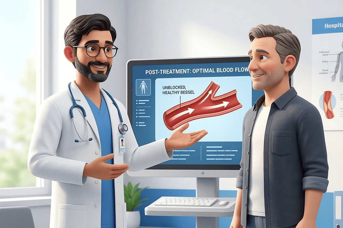
Yes, it is highly effective for treating chylothorax, especially when conservative treatments fail. Many patients experience significant improvement after the procedure.
In most cases, yes. These minimally invasive techniques avoid open chest surgery and are associated with faster recovery and fewer complications.
Recovery is usually rapid, with many patients resuming normal activities within days, depending on the severity of the lymphatic disorder.
With a commitment to patient comfort, our clinic combines cutting-edge interventional radiology with compassionate care.
connect@drsudhirvasistha.com
Copyright © 2026 All Rights Reserved. Dr. Sudhir Vasistha | Design, Developed & Powered by Better Choice