Image-Guided Biopsy Fine Needle Aspiration (FNA) & Core (CNB) for Liver, Kidney, Lung, Lymph Node, Bone, Soft Tissue, Thyroid, and Breast
General Image-Guided Interventional Procedures | Interventional Non-Vascular Procedures | Needle Biopsy | Age: Adults
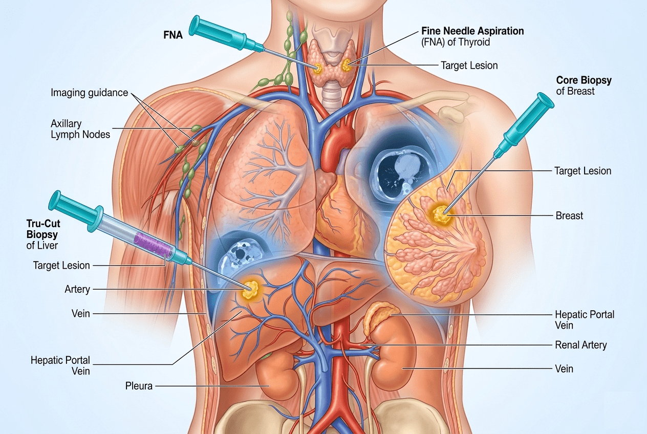
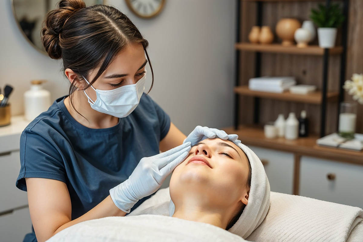
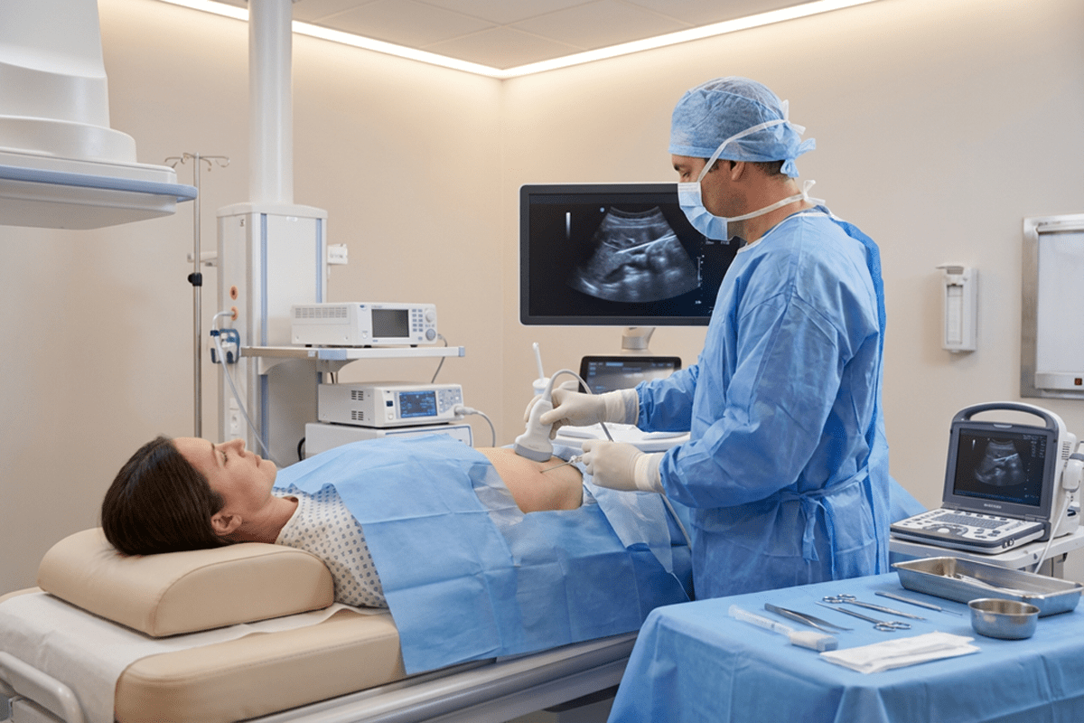
Image-guided biopsy using Fine Needle Aspiration (FNA) and Core Needle Biopsy (CNB) is a minimally invasive diagnostic procedure that allows physicians to obtain tissue samples from organs or masses with high precision. Using real-time imaging such as ultrasound, CT, or fluoroscopy, the needle is accurately guided to the target area, improving diagnostic accuracy while minimizing damage to surrounding tissue. This technique is widely used in interventional radiology for safe and reliable tissue diagnosis.
FNA collects cells for cytology, while CNB removes a small core of tissue for histology, offering more detailed analysis when needed. These outpatient procedures help diagnose cancer, infections, and inflammatory conditions while reducing the need for open surgical biopsy.
Internal links: General Image-Guided Interventional Procedures, Interventional Non-Vascular Procedures, Needle Biopsy
Image-guided biopsy is considered very safe, especially when performed by experienced interventional radiologists. Minor risks include bruising, mild pain, or bleeding at the biopsy site. Serious complications are uncommon, and every precaution is taken to ensure patient safety and comfort.
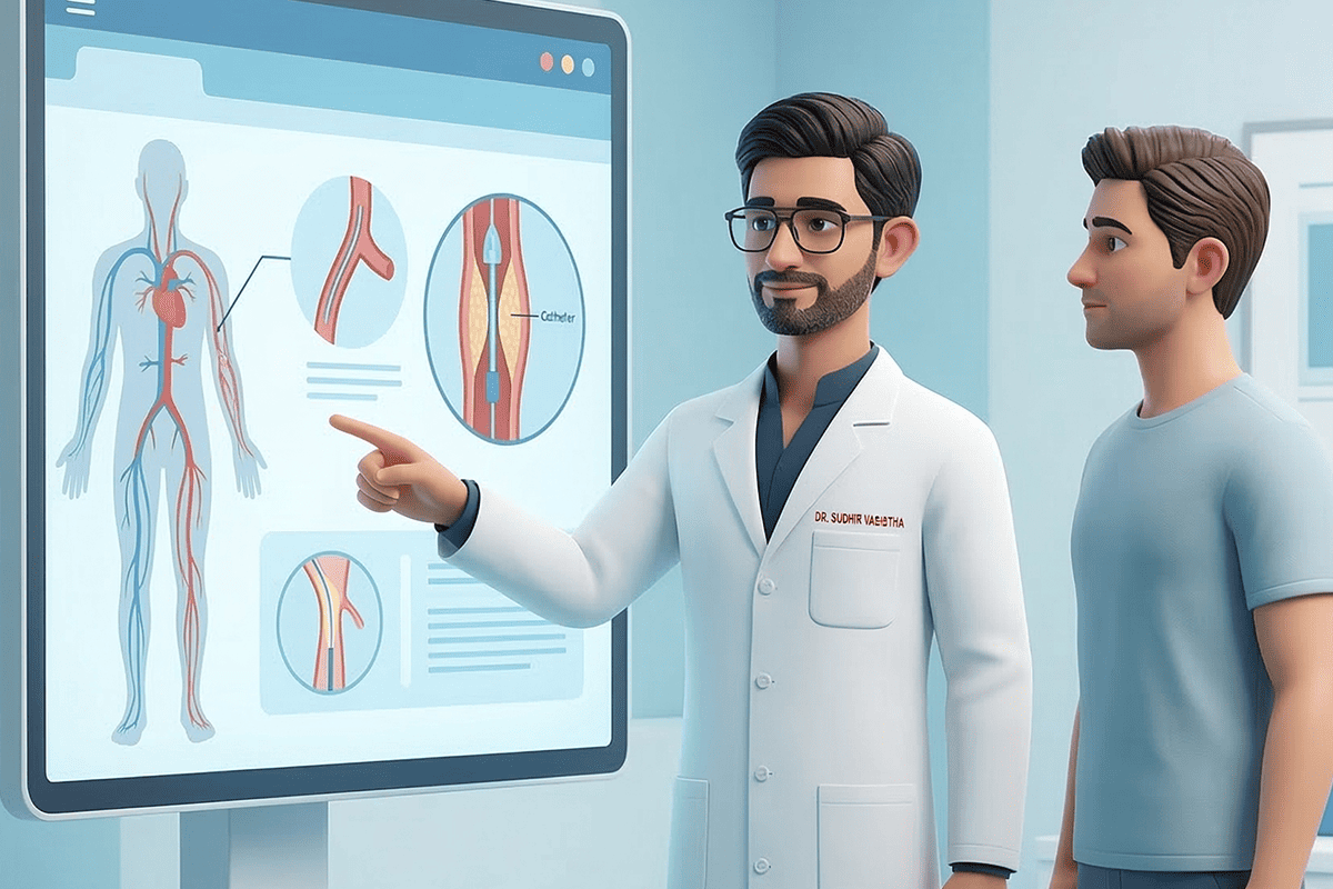
FNA removes cells for microscopic evaluation, while a core biopsy removes a small tissue sample. Your physician selects the method based on the type and location of the lesion.
Most patients experience minimal discomfort. Local anesthesia significantly reduces pain, and the procedure is generally well tolerated.
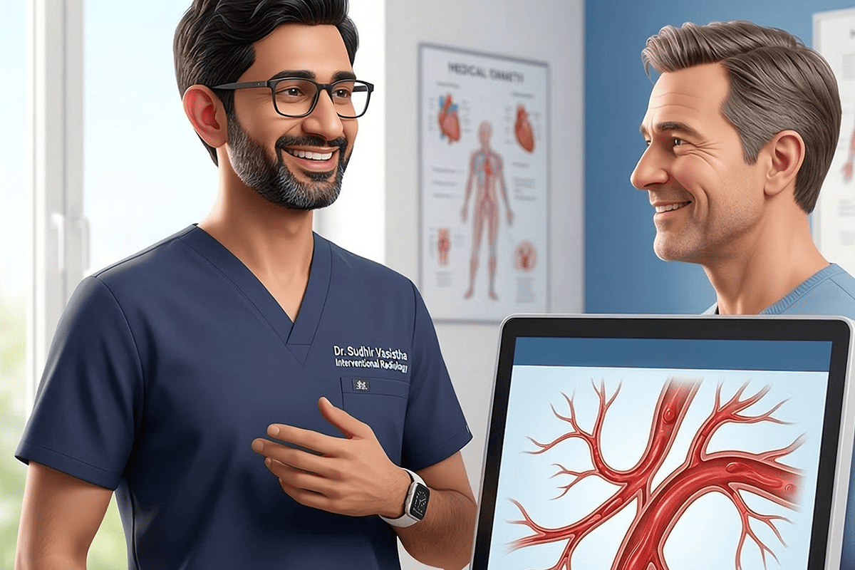
Results are typically available within a few days, depending on the complexity of testing required. Your provider will review findings and next steps with you.
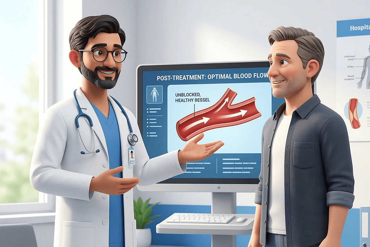
Yes, in many cases. Image-guided biopsy avoids large incisions, has fewer risks, and allows faster recovery compared to surgical biopsy.
With a commitment to patient comfort, our clinic combines cutting-edge interventional radiology with compassionate care.
connect@drsudhirvasistha.com
Copyright © 2026 All Rights Reserved. Dr. Sudhir Vasistha | Design, Developed & Powered by Better Choice